News and events
‘Twinkling Toxoplasma’ reveal potential drug targets for the treatment of parasitic infections
Winners of our third Festive Science Image Competition, run in partnership with the Medical Research Council (MRC), have been announced today.
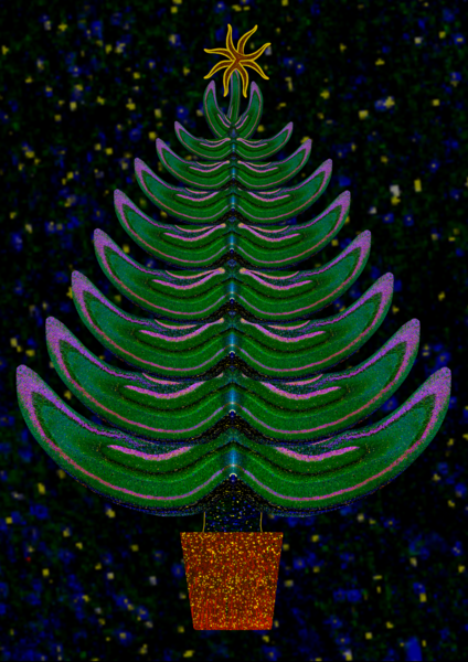
The 1st place image – ‘Twinkling Toxoplasma’ – shows human skin cells grown in a lab environment, infected with a parasite, Toxoplasma gondii. The parasites’ basket-like skeleton structures, shown in various hues, reflect the appearance of a traditional Christmas tree. This technique has the potential to illuminate otherwise unseen elements of parasites, so that areas of infection within the body can be more effectively treated.
This annual competition invites Foundation and MRC-funded researchers, staff, and students to produce a science image with direct relevance to medical research, combined with a festive theme. The competition’s judges, who work in science, medical research, communications, and public engagement, were looking for eye-catching, high-quality images, along with a clear explanation for non-scientific audiences.
Three winners were selected, with the 1st place image chosen to feature on the Medical Research Foundation and MRC's joint Season's Greetings card for 2024. The cards can be ordered online, and a suggested donation to the Foundation can be made at checkout.
Donate today
Make a difference by support life-changing research into overlooked and underfunded health conditions.
Support us
1st place – “Twinkling Toxoplasma”, by Dr Kseniia Bondarenko, Postdoctoral Research Associate from Young Lab, University of Edinburgh
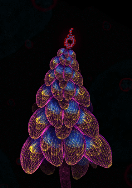
“Toxoplasmosis is an illness caused by an infection with the parasite Toxoplasma gondii. Scientists often use microscopy to explore the biology of parasites such as this, seeking clues for potential drug targets,” explains Kseniia.
To create her image, Kseniia grew human skin cells in the lab, and then infected them with Toxoplasma. She then used fluorescent dyes to highlight various parasite structures, and captured high-resolution images.
“The high-resolution images revealed the parasites’ basket-like skeleton (shown in yellow), inner shell (magenta), and DNA (blue). The ‘star’ on top of the tree is a regular-microscopy snapshot of an unusually large parasite rosette – a cluster of parasites that clump together inside the body.
“This technique has the potential to illuminate complex parasite structures, so that areas of infection within the body can be more effectively targeted and treated.”
Support us
Order cards featuring the winning image
Order your Season's Greetings cards featuring the 'Twinkling Toxoplasma' design, and make a suggested donation to the Foundation.
Order now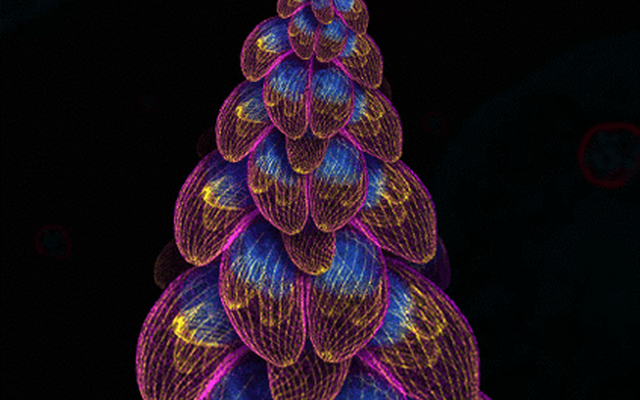
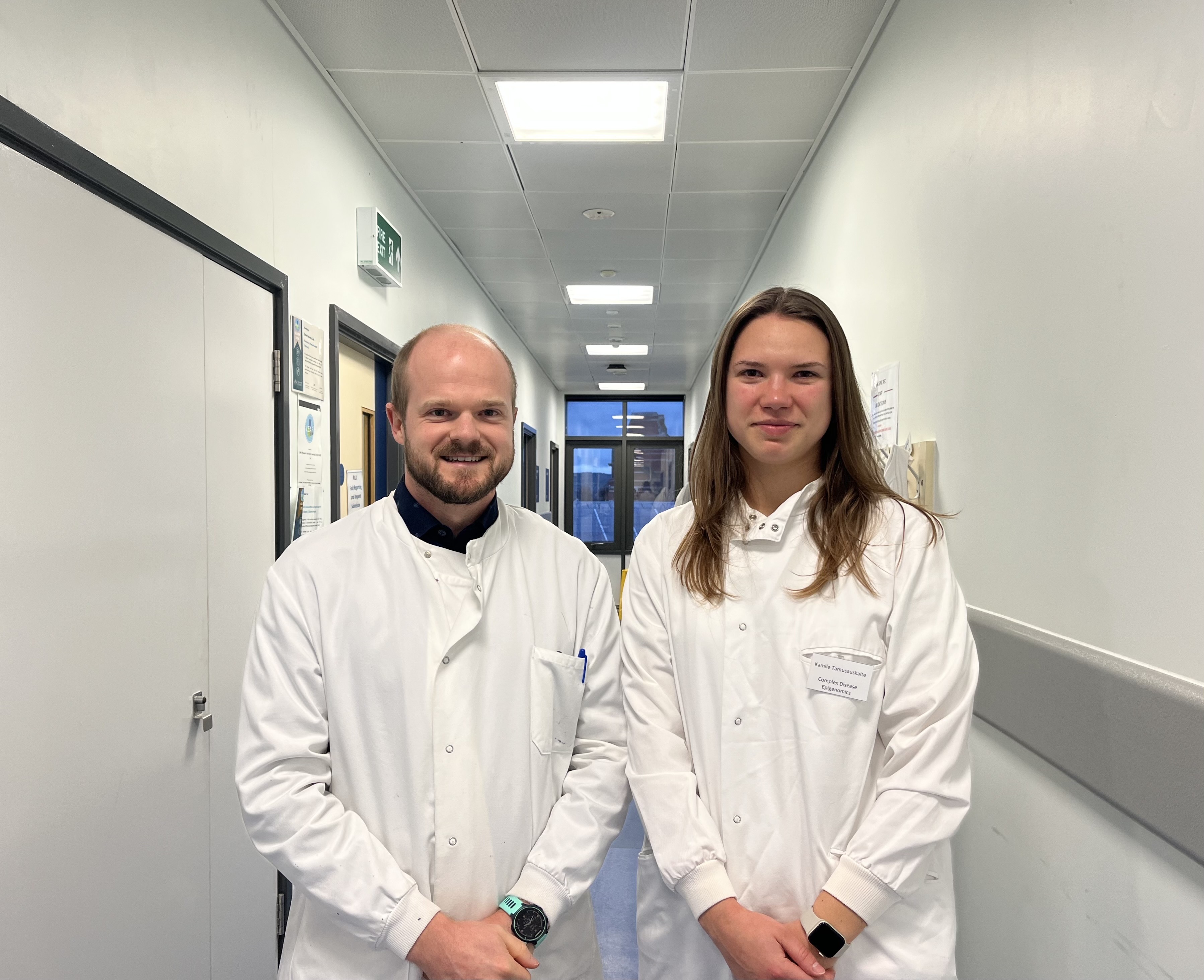
2nd place – “Christmas Memories”, by Dr Nicholas Clifton, CDA Fellow, University of Exeter & Kamile Tamusauskaite, PhD Student in Psychiatric Genomics, University of Exeter
“This image shows a section from the mouse hippocampus, a brain structure essential for forming and organising memories, like recalling last Christmas,” says Nicholas Clifton.
“The hippocampus is affected in patients with schizophrenia. We use mouse models to explore how genes linked to schizophrenia affect the hippocampus, helping us to understand the underlying biology and identify possible drug targets.
“To visualise specific proteins and their interactions in the mouse hippocampus, we used fluorescent dyes, which glow in different colours under the microscope. The resulting images highlight the classic curved shape of the hippocampus and its many layers much like a tree branch.”
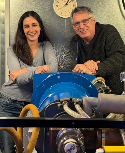
Highly commended – “Frosty Winter Mornings”, by Nathalie Lövgren, MRC DPhil Student, and Dr Iain Tullis, Senior Postdoctoral Researcher, Physics and Biology of FLASH Radiation Research Group, University of Oxford
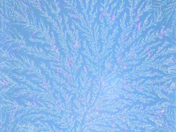
“Reminiscent of our windows during these cold winter months, this frosty scene was created by irradiating a block of Perspex using a 6-million-volt electron beam,” Nathalie and Ian explain. “The radiation charges the Perspex, and when discharged reveals the beautiful particle tracks known as ‘frozen lightning’ or a Lichtenberg figure.
“The shimmering background is FLASH radiation interacting with water, producing Cherenkov radiation, and interactions with the camera producing a pink-purple glow. Radiation therapy is a standard treatment for cancer patients today. FLASH radiotherapy is a new treatment technique which has the potential to reduce side effects commonly associated with conventional radiotherapy.”