Revealing the biology underlying human disease
Underneath all disease is biology – cells replicating out of control, organs failing, immune systems being overwhelmed, and lives being cut short. Uncovering the basic biology behind human health is essential to finding new ways to diagnose, prevent, and treat diseases better.
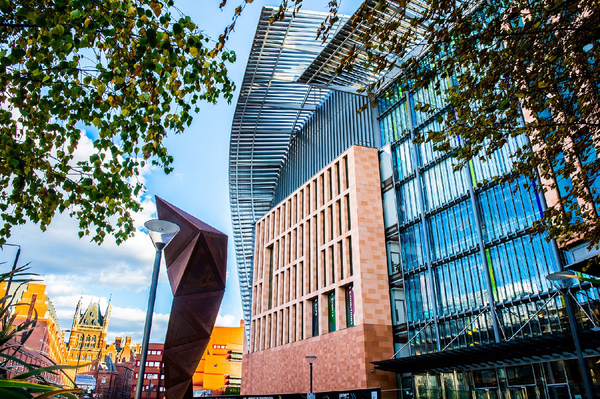
This has been the philosophy from day one of the Francis Crick Institute, a huge research lab just around the corner from London’s St Pancras Station. To understand how diseases develop, the Crick performs ‘in vivo’ research to effectively ‘see inside’ living organisms to observe how well organs are functioning, and to look at the structure of cells within the body. They have a specialised In Vivo Imaging (IVI) facility, which allows researchers to non-invasively track natural processes and diseases in live animals and monitor them over time, including ultrasound, fluorescence and Magnetic Resonance Imaging (MRI).

Dr Bernard Siow is the head of MRI in the IVI facility. Bernard says that, in the same way that a lab scientist would use a microscope, in vivo imaging is like a microscope that reveals the details of how diseases develop inside living beings.
Cutting-edge equipment
Set up with our help, through a £1.8 million donation, the IVI facility houses the Crick’s cutting-edge imaging equipment and a team of experts to help scientists use it. As well as scientific imaging equipment, there are miniature versions of hospital scanners including CT, MRI, and PET, as well as ultrasound machines.
Crick scientists are using the IVI facility in lots of different ways. Sometimes the equipment is used for relatively basic but important functions, such as accurately measuring the size of tumours to track how cancer grows.
Other projects involve Bernard and the IVI team developing bespoke imaging techniques for researchers. And the benefit of having hospital style research scanners is that these new imaging techniques could potentially be used for human patients in the future.
The IVI facility has significantly improved how animal research is carried out at the Crick. For example, in vivo imaging allows scientists to study diseases inside mice without needing to sacrifice and dissect them, which reduces the numbers of mice used in experiments and the harm they might experience. And studying a disease in one mouse over time, rather than in different mice at different stages, can give a better picture of how the disease really develops.
Part of our donation paid for a single-photon emission computed tomography (SPECT) scanner. This provides scientists with 3D images of blood flow to show how certain molecules are being used and how they travel through the animals’ bodies. One research team is using the SPECT scanner to monitor the growth and spread of tumours from cancer cells in mice.
Without that contribution [from the Foundation], the IVI facility would be much smaller, would have far fewer machines, and would be far less rounded. Now I would say we are one of the top imaging facilities in the UK. Dr Bernard Siow, Francis Crick Institute

A central hub of expertise
But the equipment is not the IVI facility’s only asset. Having everything in one place with the support of experts like Bernard is a key strength. This allows the scientists to get the most out of the facility and the best from their research. “Bringing everything together really just helps to centralise things, so that people feel comfortable to come to us, exchange ideas with other experts from different fields, and use the equipment,” says Bernard.
The IVI facility is making a big contribution to the research at the Crick, and Bernard believes our support has been crucial to its success. “I don’t think we would have been able to create such a facility without the Foundation’s donation,” he says.
“Without that contribution, the IVI facility would be much smaller, would have far fewer machines, and would be far less rounded. Now I would say we are one of the top imaging facilities in the UK.”
Thank you to GlaxoSmithKline, who made the donation to the Crick’s IVI facility possible.


