Imaging progress at the Francis Crick Institute
Our funding is helping Crick scientists visualise and track disease in living animals.
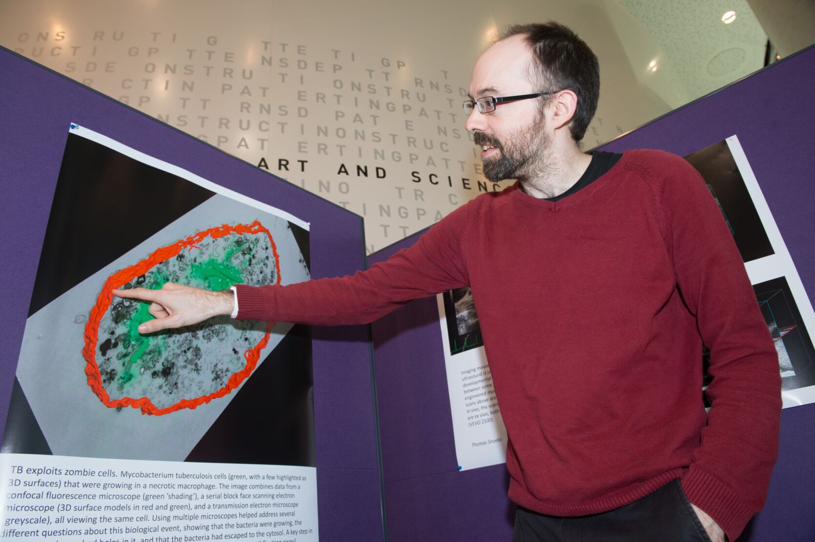
Eighteen months ago, the Francis Crick Institute’s biological imaging facility, funded by a grant from the Medical Research Foundation, opened to researchers.
The in vivo imaging project is a perfect example of how Medical Research Foundation funding helps scientists push ahead with their research ambitions. By investing directly in imaging facilities at The Francis Crick Institute we have enhanced the facilities available to scientists there and nurtured research ideas that have fantastic potential to improve people’s health and lives.Angela Hind
Chief Executive

Its powerful visualisation systems let scientists combine molecular, functional and structural images to build a complete picture of disease. The facility is supported by a £1.8million grant from the Medical Research Foundation, made possible thanks to a donation from GlaxoSmithKline (GSK).
Yesterday the Foundation met to explore work the imaging facility has made possible so far. Since it opened in January 2017, scientists from many research fields have made use of the bio-imaging equipment to help answer questions about how and why disease develops. Their answers should help find new ways to prevent, diagnose or treat illnesses including cancer, heart disease, stroke, infection and neurodegeneration.
The facility is equipped with SPECT/CT and PET/MRI systems, as well as a 9.4T preclinical MRI system. These powerful tools capture high-resolution images inside living animals so that scientists can monitor changes to health and disease over time.
Diana Passaro, a scientist working on the project said: “We’re starting by imaging mice, but if our findings are promising, it could have direct implications for diagnostic and prognostic approaches in human patients.”
The facility is also home to optical imaging systems including IVIS Spectrum fluorescence / bioluminescence imagers, a Pearl NIR imager, an intra-vital microscope, and two dedicated small animal high-resolution ultrasound machines.
Having multiple types of imaging equipment in one space enables scientists to measure different things in a single animal within a short time frame. This is particularly useful when multiple techniques are required to see the whole picture. For example, neuroscientists trying to work out which parts of the brain are stimulated by a set of electrodes can use X-ray CT imaging to show where exactly the electrodes are located in the brain, and MRI imaging to reveal the corresponding brain activity.
“It’s amazing to see how, in a relatively short space of time, the facility has got up-and-running and is being widely used by Crick scientists and their collaborators to make new research discoveries,” Bernard Siow, Head of the MRI facility.